Dental plaque is not random "gunk" on your teeth — it is a structured oral biofilm, a highly organized microbial community that forms in predictable stages. Within minutes of cleaning your teeth, a protein film (pellicle) forms on enamel, and pioneer bacteria begin colonizing within hours. Over days, the biofilm matures into a complex, three-dimensional structure that becomes increasingly resistant to removal and antimicrobials. Understanding how biofilm develops — and the specific bacteria involved at each stage — explains why consistent, thorough oral hygiene matters so much more than occasional deep cleaning.
If you have ever run your tongue across your teeth several hours after brushing and felt a fuzzy film, you have encountered oral biofilm firsthand. That slippery coating is not just food residue — it is a living, organized community of bacteria embedded in a self-produced matrix. Dental plaque, the clinical term most people know, is simply what this biofilm looks and feels like on tooth surfaces.
What makes oral biofilm fascinating — and clinically significant — is that it is not a random accumulation. It develops through highly ordered stages, involves specific bacterial species at each step, and becomes progressively harder to remove as it matures. Research over the past three decades has transformed our understanding of plaque from "dirty stuff on teeth" to "one of the most studied biofilm systems in biology."
What Is a Biofilm?
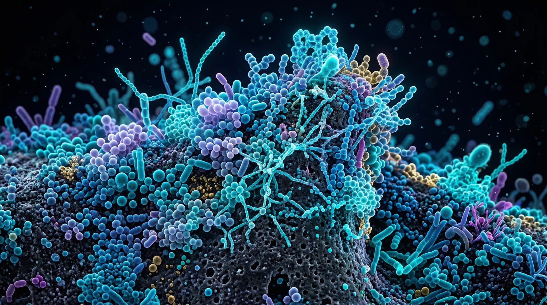
A biofilm is any community of microorganisms that attaches to a surface and encases itself in a self-produced matrix of extracellular polymeric substances (EPS) — a sticky mixture of polysaccharides, proteins, lipids, and extracellular DNA. Biofilms exist throughout nature: on rocks in streams, inside water pipes, on medical implants, and on virtually every moist surface in the human body.
The oral biofilm that forms on teeth is one of the most diverse biofilm systems known. The human mouth harbors approximately 700 identified bacterial species, and a single milliliter of saliva may contain over 100 million bacterial cells. A mature dental plaque biofilm can contain more than 200 different species organized in spatial relationships that facilitate cooperation, communication, and collective survival.
What makes biofilms fundamentally different from free-floating (planktonic) bacteria is their structure. Bacteria within a biofilm are embedded in the EPS matrix, which acts as a physical barrier against antimicrobial agents, host immune defenses, and mechanical removal. Research published in Clinical Microbiology Reviews, 2002 demonstrated that bacteria within biofilms can be up to 1,000 times more resistant to antibiotics than the same species in planktonic form.
This is why dental plaque is so difficult to eliminate with mouthwash alone — and why physical disruption through brushing and flossing remains essential.
How Dental Plaque Forms: The Five Stages
Plaque formation follows a well-characterized developmental sequence that researchers have documented through decades of clinical observation and advanced imaging techniques.
Stage 1: Pellicle Formation (0-2 Minutes)
Within seconds of cleaning your teeth, a thin protein film called the acquired enamel pellicle begins forming on tooth surfaces. This pellicle is derived from salivary glycoproteins, phosphoproteins, and enzymes that selectively adsorb onto the hydroxyapatite crystal structure of enamel.
The pellicle is not bacterial — it is an acellular protein layer approximately 0.1 to 1 micrometer thick. However, it plays a critical role in biofilm development because it modifies the tooth surface chemistry, creating specific binding sites (receptors) that certain bacterial species can recognize and attach to.
A study in Advances in Dental Research, 1997 identified over 130 different proteins in the acquired pellicle, many of which serve as receptors for specific bacterial adhesins (attachment molecules). The pellicle essentially "prepares" the tooth surface for bacterial colonization.
Stage 2: Early Colonization (0-4 Hours)
The first bacteria to attach to the pellicle-coated tooth surface are called primary or pioneer colonizers. These are predominantly gram-positive, aerobic or facultatively anaerobic species that can tolerate the oxygen-rich environment near the tooth surface.
The key pioneer colonizers include:
- Streptococcus sanguinis — One of the most abundant early colonizers, considered a health-associated species that competes with cariogenic bacteria
- Streptococcus gordonii — Produces hydrogen peroxide, which may inhibit some pathogenic species
- Streptococcus mitis — A commensal species well-adapted to oral mucosal and enamel surfaces
- Actinomyces naeslundii — Attaches to pellicle proteins and co-aggregates with streptococcal species
These pioneer species attach through specific molecular interactions — their surface adhesins bind to complementary receptors in the pellicle. This specificity explains why the same species consistently appear as first colonizers across different individuals.
Research in the Journal of Bacteriology, 2005 demonstrated that early colonizers express multiple adhesins allowing them to bind to different pellicle components, giving them a competitive advantage in the initial colonization race.
Stage 3: Co-Aggregation and Diversification (4-24 Hours)
Once pioneer species establish themselves, they create conditions that allow secondary colonizers to attach — not to the tooth surface directly, but to the pioneer bacteria themselves. This process is called co-aggregation: specific cell-to-cell recognition between different bacterial species.
The most important bridge organism in this process is Fusobacterium nucleatum. Research published in Periodontology 2000, 2005 demonstrated that F. nucleatum can co-aggregate with virtually every other oral species tested — both early and late colonizers. It literally bridges the gap between the health-associated pioneer community and the pathogenic species that arrive later.
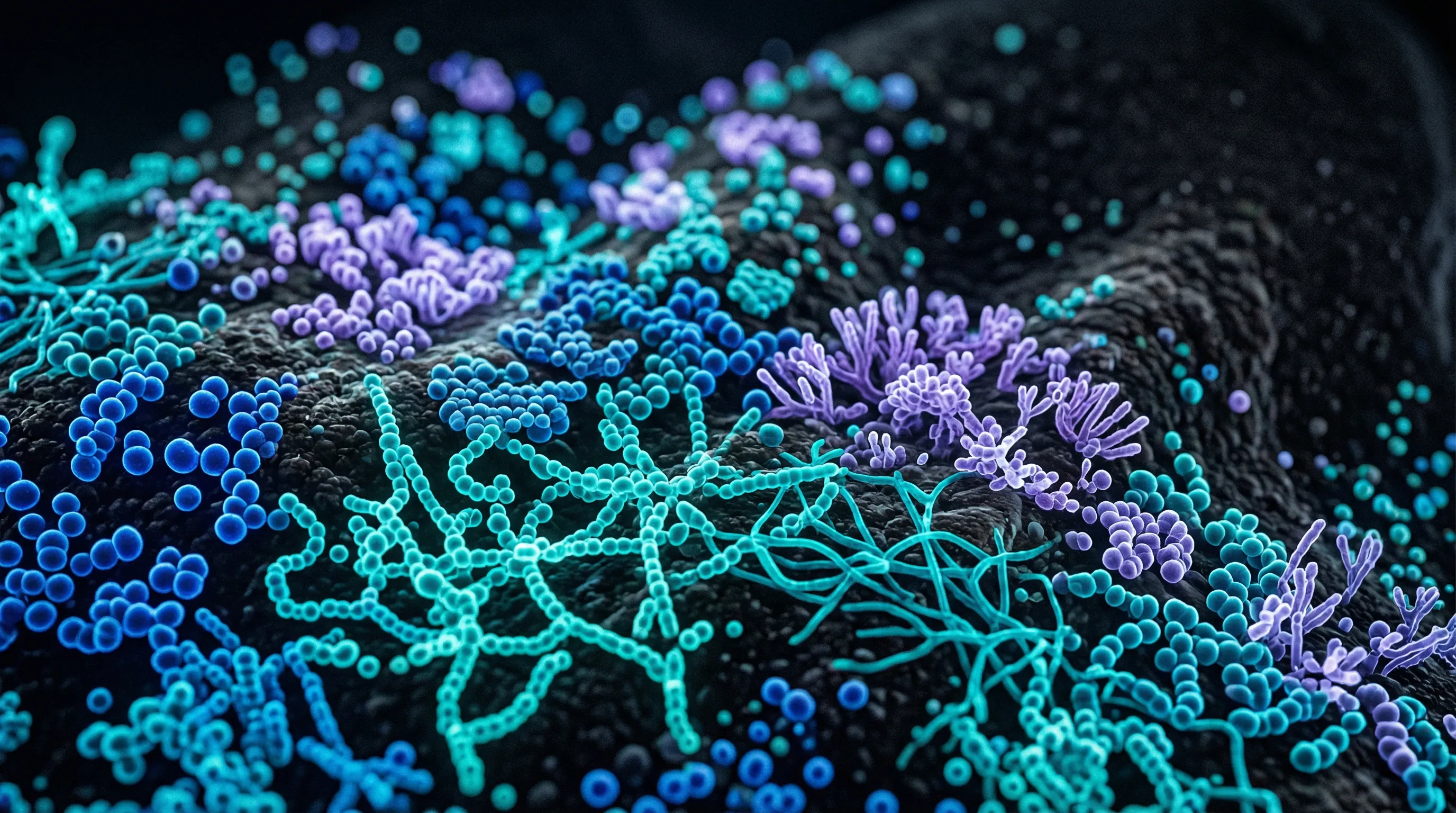
During this stage, the biofilm begins developing its three-dimensional architecture. Bacteria produce EPS, creating water channels that allow nutrient transport to deeper layers and waste removal from the interior. The community transitions from a thin monolayer to a structured, multi-species consortium.
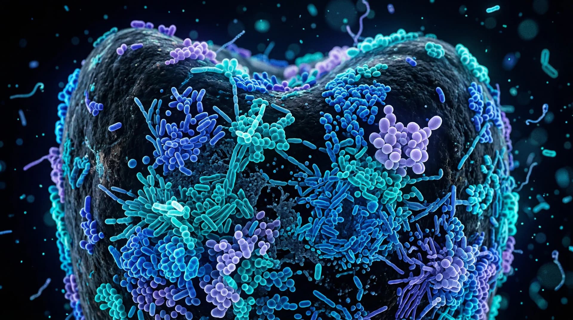
Stage 4: Mature Biofilm (1-14 Days)
As the biofilm matures, its composition shifts dramatically. Oxygen consumption by surface bacteria creates anaerobic zones in deeper layers, allowing obligate anaerobes to establish themselves. The species that appear at this stage are often the ones associated with dental disease:
- Streptococcus mutans — The primary bacterium associated with tooth decay; produces copious lactic acid from sugar fermentation and thrives in the acidic conditions it creates
- Porphyromonas gingivalis — A keystone pathogen in periodontal disease; manipulates the host immune response and disrupts microbial community balance
- Treponema denticola — A spirochete associated with advanced periodontal disease
- Tannerella forsythia — Part of the "red complex" of periodontal pathogens alongside P. gingivalis and T. denticola
A landmark study published in Microbiology and Molecular Biology Reviews, 2000 by Socransky and colleagues classified oral bacteria into color-coded complexes based on their association patterns. The "red complex" (P. gingivalis, T. denticola, T. forsythia) was most strongly associated with deep periodontal pockets and clinical signs of disease.
The mature biofilm is now a formidable structure — organized, cooperative, and resistant. Bacteria within it communicate through quorum sensing molecules, share nutrients, transfer antibiotic resistance genes, and collectively resist both the host immune system and antimicrobial treatments.
Stage 5: Calcification — From Plaque to Tartar (Days to Weeks)
If plaque remains undisturbed, minerals from saliva (primarily calcium and phosphate) gradually deposit within the biofilm matrix, hardening it into dental calculus (tartar). Calculus cannot be removed by brushing or flossing — it requires professional scaling by a dentist or hygienist.
Calculus itself is not directly pathogenic, but its rough surface provides an ideal substrate for new biofilm attachment, perpetuating the cycle. Subgingival calculus (below the gumline) is particularly problematic because it harbors anaerobic pathogens in close proximity to gum tissue, contributing to gingivitis and periodontitis progression.
S. Mutans and P. Gingivalis: Two Key Players
Understanding two bacteria in particular helps illustrate how biofilm composition determines disease outcomes.
Streptococcus mutans is the most studied cariogenic (cavity-causing) bacterium. It possesses several properties that make it uniquely destructive within the biofilm: it metabolizes sucrose into lactic acid more efficiently than most oral bacteria, it produces sticky glucans (from sucrose via the enzyme glucosyltransferase) that strengthen its attachment within the biofilm, and it thrives at low pH levels that kill competing species — a phenomenon called aciduricity. As S. mutans dominates, the local environment becomes more acidic, favoring further S. mutans growth while suppressing health-associated species. This self-reinforcing cycle is what drives tooth decay through its progressive stages.
Porphyromonas gingivalis, by contrast, is a periodontal pathogen rather than a cariogenic one. Present in small numbers in many healthy mouths, P. gingivalis becomes destructive when it reaches sufficient abundance to manipulate the host immune response. Research in Cell Host & Microbe, 2012 introduced the concept of P. gingivalis as a "keystone pathogen" — a species that, even at low abundance, can restructure the entire microbial community toward dysbiosis. It subverts the host immune response in ways that increase inflammation while impairing bacterial clearance, creating an environment where other pathogens flourish. The resulting chronic inflammation and tissue destruction is what causes bleeding gums, pocket deepening, and eventual bone loss in periodontal disease.
The Good Biofilm vs. Bad Biofilm Debate
A common misconception is that all dental plaque is harmful and should be eliminated entirely. The reality is more nuanced. The early-stage biofilm dominated by health-associated species (S. sanguinis, S. gordonii, Actinomyces species) is not inherently pathogenic — in fact, these bacteria may be protective.
Research suggests that a balanced oral biofilm serves several beneficial functions:
- Colonization resistance — Health-associated bacteria occupy ecological niches, preventing pathogenic species from establishing themselves
- Immune modulation — Commensal bacteria "train" the local immune system to respond appropriately to genuine threats
- Mineral homeostasis — Some biofilm bacteria help maintain the balance between demineralization and remineralization on enamel surfaces
The problem is not biofilm per se — it is uncontrolled biofilm maturation. When plaque is allowed to accumulate undisturbed for days, the ecological succession described above shifts the community toward pathogenic dominance. The goal of oral hygiene is not to sterilize the mouth (which is impossible and undesirable) but to regularly reset the biofilm to its early, health-associated state.
This perspective explains why aggressive antiseptic mouthwash use can sometimes backfire — by killing bacteria indiscriminately, it may eliminate the beneficial species that provide colonization resistance, allowing pathogens to recolonize first.
How to Effectively Disrupt Biofilm
Understanding biofilm biology informs practical oral hygiene strategies:
Mechanical disruption is non-negotiable. Because of the EPS matrix, chemical agents alone cannot adequately penetrate mature biofilm. Brushing twice daily with a soft-bristled brush physically disrupts the biofilm structure, exposing bacteria to saliva, oxygen, and antimicrobial agents. Electric toothbrushes may provide superior biofilm removal compared to manual brushing, according to a Cochrane review published in Cochrane Database of Systematic Reviews, 2014.
Interdental cleaning reaches surfaces brushing misses. Approximately 30% of tooth surface area is interdental — accessible only by floss, interdental brushes, or water flossers. Biofilm in these areas matures undisturbed unless specifically targeted, which is why people who brush well but do not floss may still develop interproximal cavities and gum disease.
Timing matters more than intensity. Because biofilm takes roughly 24 hours to mature beyond the pioneer stage, disrupting it once or twice daily is sufficient to prevent pathogenic succession. You do not need to brush harder — you need to brush consistently and thoroughly. Consult your dentist if you are unsure whether your technique adequately disrupts biofilm in all areas.
Diet affects biofilm composition. Frequent sugar consumption fuels S. mutans acid production and glucan synthesis within the biofilm, accelerating its shift toward cariogenic dominance. Eating foods that support gum health and reducing sugar frequency may help maintain a healthier biofilm ecology.
ProDentim
Oral Probiotic for Gum & Teeth Health
ProDentim delivers beneficial bacterial strains — including L. reuteri and L. paracasei — directly to the oral cavity via a dissolving tablet. The goal is to support the colonization of health-associated species that compete with pathogenic biofilm inhabitants like S. mutans and P. gingivalis.
We may earn a commission if you make a purchase through our links, at no extra cost to you.
The Clinical Bottom Line
Dental plaque is a biofilm — a structured, cooperative microbial community that develops in predictable stages on tooth surfaces. Its early stages are dominated by health-associated species, but without regular disruption, it matures into a pathogenic community capable of causing both cavities and gum disease. The transition from health-associated to pathogenic biofilm is driven by ecological succession, with bridge organisms like F. nucleatum facilitating the arrival of destructive species.
Effective oral care is fundamentally about biofilm management: disrupting the community regularly through mechanical cleaning, supporting beneficial species through balanced nutrition and targeted probiotics, and preventing the accumulation that leads to calculus formation. Regular professional cleanings complement daily home care by removing calcified deposits that home hygiene cannot address.
This article is for educational purposes only and does not constitute medical advice. Always consult your dentist or healthcare provider for personalized guidance on oral hygiene and disease prevention.